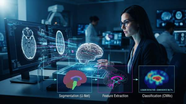
Artificial intelligence in medical imaging is best understood not as a single clever algorithm but as a sequence of computational stages that carry a raw scan from acquisition to diagnostic insight. A survey of AI applications in medical imaging describes this shift as a move away from conventional step-by-step manual interpretation toward a data-driven pipeline in which models perform automated preprocessing, feature extraction, and organized reporting. This article walks through that pipeline stage by stage, identifying which algorithm types—such as convolutional neural networks for classification or U-Net-style architectures for segmentation—do the work at each point.
Introduction: The AI Medical Imaging Analysis Pipeline
The pipeline model matters because it explains why so many different techniques coexist under the banner of "AI in imaging." Digital medical images arrive in many modalities—ultrasound, X-ray, CT, MRI, PET, retinal photography, histology slides, and dermoscopy images, as an Amazon Web Services technical overview of medical image analysis pipelines notes. No single algorithm handles all of these equally well; instead, different stages of analysis call for different computational approaches.
One important caveat frames everything that follows: this pipeline is a conceptual model synthesized from descriptions of various AI applications in medical imaging, not a single, universally defined standard. Different systems may merge, reorder, or skip stages. But the four-stage structure—preprocessing, segmentation, feature extraction, and classification—captures how these systems are typically described in the literature and in practice.
The AI Medical Imaging Analysis Pipeline
The table below deconstructs the end-to-end process into its functional stages, mapping each to its purpose, the algorithm types associated with it, and a representative application. Together, the rows let you trace the path from a raw medical image to a diagnostic insight.
| Stage | Purpose | Illustrative Algorithms | Example Application |
|---|---|---|---|
| Image Preprocessing | Prepare raw images for analysis; AI models perform automated preprocessing operations and image-quality enhancement | Deep learning-based quality enhancement | Improving image quality and reducing scanning time in liver and pancreatic imaging |
| Image Segmentation | Isolate regions of interest such as organs, lesions, or tumors | U-Net and Attention U-Net architectures | MRI segmentation; PET head motion estimation research at Yale's biomedical imaging institute |
| Feature Extraction | Identify and quantify hidden patterns that humans cannot detect | Radiomics-driven deep learning; dimensionality-reduction techniques | Extracting quantitative parameters such as lesion volume and tumor burden |
| Classification / Prediction | Classify images, detect anomalies, and forecast disease presence and progression | Convolutional neural networks (CNNs); predictive analytics models | Diabetic retinopathy screening from retinal images |
Reading down the "Purpose" column shows the logic of the sequence: each stage reduces the problem for the next. Preprocessing makes the data tractable, segmentation narrows attention to relevant anatomy, feature extraction converts pixels into measurable quantities, and classification turns those quantities into a diagnostic statement or risk estimate.
Stage 1: Image Preprocessing and Data Preparation
Preprocessing is the stage where raw images become usable inputs. According to the clinical radiology literature, AI models perform automated preprocessing operations and contribute to image-quality enhancement before deeper analysis begins. This stage exists because medical imaging data is complex and heterogeneous across modalities, and deep learning methods must contend with that complexity before patterns can be learned reliably.
The practical value of automated preprocessing is already visible in real workflows. A survey of AI in medical imaging notes that for liver and pancreatic images, AI algorithms are already speeding up repetitive tasks, improving image quality, reducing scanning time, and optimizing image acquisition. In other words, the first stage of the pipeline is not merely a technical prerequisite—it can directly affect the patient experience by shortening scans.
Stage 2: Image Segmentation for Region Isolation
Segmentation answers the question: where in the image is the relevant anatomy? Algorithms at this stage draw boundaries around organs, lesions, or tumors so that subsequent analysis focuses on the right structures. This is where U-Net-style architectures come in. The Yale Biomedical Imaging Institute's Image Analysis/AI Interest Group, whose members develop and use these methods, lists Attention U-Net among the algorithm families its researchers work with, alongside supervised deep learning for PET motion estimation and MRI segmentation.
Segmentation is also where AI delivers some of its most tangible quantitative benefits. In liver and pancreatic imaging, the survey literature reports that AI-driven segmentation enables the acquisition of new quantitative parameters such as lesion volume and tumor burden—measurements that are repetitive and time-consuming to produce manually. By automating the delineation step, the pipeline converts an image into a set of numbers that clinicians can track over time.
Stage 3: Feature Extraction and Representation
Once a region of interest is isolated, feature extraction quantifies its relevant characteristics. The clinical radiology literature describes deep learning methods leveraging radiomics to extract hidden patterns from CT, MRI, and PET images that cannot be detected by humans. This is a distinctive claim worth pausing on: the value of this stage is not just speed, but the ability to surface signal that a human reader would never see.
Feature extraction also addresses a fundamental computational problem—data complexity. Deep learning methods associated with this stage employ dimensionality-reduction techniques to manage the sheer volume of information in medical images, distilling thousands of pixel values into a compact representation that a classifier can work with. The qualification matters here: the evidence links dimensionality reduction to deep learning methods and feature extraction broadly, rather than to preprocessing specifically.
Stage 4: Classification and Predictive Modeling
The final stage converts extracted features into answers. Convolutional neural networks are the workhorse for classification tasks—Spectral AI, a medical imaging company, describes modern AI systems using CNNs, generative models, and segmentation algorithms to detect anomalies, classify tissues, and support diagnostic decisions. The same clinical radiology literature describes this stage as encompassing anomaly detection and predictive modeling, with AI models generating organized reports that present results through detailed analysis.
Predictive modeling extends beyond the image itself. AI models can integrate imaging data with patient history, biomarkers, and demographic factors to forecast risks and outcomes, according to Spectral AI's description of predictive analytics. This is where the pipeline crosses from detection into prognosis: not just "is there a lesion" but "what is likely to happen to this patient."
Applications: Detecting Diseases Across Modalities
The pipeline's stages come together in disease-specific applications. In cancer detection, AI algorithms applied to X-rays, CT, MRI, and ultrasound can identify subtle abnormalities, including oncologic changes, that may be difficult to detect visually. In neurodegenerative disorders, the clinical radiology literature describes AI applied to medical imaging for diagnosis and assessment of these conditions, analyzing complex data associated with them.
Diabetic retinopathy offers perhaps the clearest end-to-end example. A survey of automated retinal disease assessment describes AI helping healthcare workers with early detection, screening, diagnosis, and grading of retinal diseases including diabetic retinopathy, retinopathy of prematurity, and age-related macular degeneration. Deep learning and CNNs specifically have been applied to retinal image analysis for diabetic retinopathy screening—a disease where early detection and intervention can change outcomes, as it is a significant cause of blindness globally.
Transparency is an emerging consideration across all these applications. The clinical radiology literature notes that tools helping doctors understand the causes of model-based lesion or anomaly detection fulfill needs for both diagnostic confidence and transparency—suggesting that the pipeline's outputs must be explainable, not just accurate, to earn clinical trust.
Enhancing Diagnostic Capabilities with AI
Clinicians and researchers should recognize the structured, multi-stage nature of AI in medical imaging to effectively integrate and develop these technologies for improved diagnostics. Understanding that preprocessing, segmentation, feature extraction, and classification are distinct stages with distinct algorithm families makes it easier to evaluate where a given tool fits, what data it depends on, and where its outputs should be checked.
Two boundaries keep this picture honest. The algorithms named here—CNNs, U-Net, Attention U-Net, radiomics-driven deep learning—are illustrative examples, not an exhaustive catalog. And this article does not evaluate the clinical efficacy or regulatory approval status of any specific tool. The measurable indicator to watch is increased adoption of AI-powered diagnostic tools in clinical settings: as the survey literature notes, AI is already a reality for repetitive tasks in liver and pancreatic imaging, and the spread of that pattern into other organs and modalities is the concrete signal of the pipeline's real-world impact.
Sources
- How Artificial Intelligence Is Shaping Medical Imaging Technology: A Survey of Innovations and Applications - PMC
- Build a medical image analysis pipeline on Amazon SageMaker using the MONAI framework — Amazon Web Services
- AI in Clinical Radiology: Addressing Core Medical Imaging Diagnosis Challenges — LINK
- Analysis/AI — Yale Biomedical Imaging Institute
- Artificial Intelligence in Medical Imaging — Spectral AI